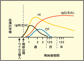
Complement fixed with antigen antibody complex is
indirectly demonstrated by the non-hemolysis of sensitized blood cells as indicators.
High group specificity
Relatively early antibody disappearance
For infection screening
Hemagglutination inhibition reaction (HI)
In the case of viruses that have hemagglutinating abilities,
antibodies that inhibit hemagglutination are demonstrated.
High type specificity
Antibodies rise early and persist
Fluorescent antibody metod (FA)
The reaction between the antibody and the viral antigen
in the infected cells is demonstrated using fluorescently labeled antibodies.
Antibody fractionation is possible.
Neutralization reaction (NT)
Active viruses are neutralized by antibodies,
proving protective antibodies against infection.
High type specificity
Enzyme immunoassay (EIA)
The antibody is reacted with the immobilized viral antigen,
and then demonstrated by its reaction with an enzyme-labeled antibody.
Antibody fractionation is possible
Quantitative data
High sensitivity compared to other methods
Chemiluminescence Immunoassay (CLIA)
This method measures the presence or amount of antibodies
by reacting antibodies in a sample with antigens immobilized
on magnetic particles and antigens labeled with chemiluminescent substances
and measuring the intensity of the resulting luminescence.
High specificity
High sensitivity
Passive (particle) agglutination reaction (PA)
Viruses are adsorbed on immobilized gelatin particles,
which are then reacted with antibodies, and proven
by the presence or absence of agglutination.
High sensitivity
Immunochromatography (IC)
This method detects antibodies that react specifically
with HIV-1 or HIV-2 antigens immobilized on a membrane filter.
High sensitivity and high specificity
Confirmation test
Characteristic of antibodies to be detected
Characteristic
Kinetics
Antiviral antibody activity
Complement fixation ability
Placental transfer
Antibodies
IgM
Produced early but disappears in a short time
+
+
-
IgG
Appears later than IgM. Persists for a long period while gradually decreasing.
+
+
+
IgA
Appears slightly later than IgM but can be detected for a longer period than IgM
+
-
-
Interpretation of antibody titer and significance of paired serum test
There is no such concept as "a normal value" for viral serum antibody titers.
Detection of antibodies produced after viral infection only retrospectively
indicates past infection with that virus and does not necessarily
reflect the current condition.
Viral antibodies are high immediately after infection and decreases thereafter,
but it is often not possible to determine whether or
not there was an infection in the recent past based only on the antibody titer
of a single serum.
It is necessary to understand the antibody response pattern after viral infection,
the characteristics of each test method, and the significance of the test,
and select the test method according to the purpose.
If the antibody titer of paired sera in the acute phase (early after the onset of illness)
and the recovery phase (14 to 21 days after the onset of illness) increases by 4-fold or more,
it is considered significant and infection with that virus is suspected.
However, an increase in antibody titer when gamma globulin is
administered for treatment is not necessarily considered significant.
Guidelines for selecting a test method according to the purpose
The test method should be selected according to the purpose depending on its characteristics.
In natural infection, it is useful to detect IgM antibodies that respond at the early stage of
infection and to observe the rise in antibodies using paired serum.
In addition, EIA for IgG antibodies is useful for determining whether there is a history
of infection and the effectiveness of vaccines.
Natural infection
Presence of history of infection
Effectiveness of vaccines
Measles
NT, EIA (IgM) (IgG)
NT, EIA (IgG)
NT, EIA (IgG)
Rubella
HI, EIA (IgM) (IgG)
HI, EIA (IgG)
HI, EIA (IgG)
Mumps
CF, NT, EIA (IgM) (IgG)
EIA (IgG)
NT, EIA (IgG)
Varicella
CF, EIA (IgM) (IgG)
EIA (IgG)
EIA (IgG)
Japanese Encephalitis
HI, CF
HI
Influenza
CF, HI
HI
Characteristics of viral antigen test
Test Method
Principle
Characteristic
Enzyme immunoassay (EIA)
The viral antigen is reacted with the specific antibody and detected by an enzyme reaction. There are two methods: the direct method, in which an enzyme is directly labeled to the specific antibody for detection, and the indirect method, in which an enzyme is labeled to the secondary antibody.
High sensitivity
Fluorescent antibody metod (FA)
The viral antigen is reacted with the specific antibody and detected using fluorescent dyes. There are two methods: the direct method, in which the specific antibody is directly labeled with a fluorescent substance for detection, and the indirect method, in which the secondary antibody is labeled with a fluorescent substance.
High specificity
Amplification of gene (PCR)
The target primer (a 20-30 base DNA fragment complimentary to each DNA end of the region to be specifically amplified) is bound to heat-denatured single-stranded DNA, and a DNA synthesis reaction is carried out using DNA polymerase. This process is repeated to exponentially amplify the target DNA sequence.
High sensitivity and high specificity
Southern blot hybridization
The sample DNA is digested with restriction enzymes and fractionated by agarose electrophoresis, and the denatured single-stranded DNA is transferred to a membrane and hybridized with a labeled probe to detect the target gene.
Analysis of quantitative and qualitative abnormalities in DNA